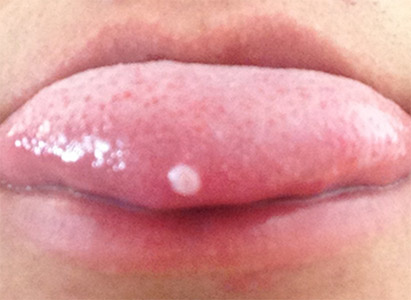
Tongue cancer is a malignant tumor occurring in the tongue, divided into oral tongue carcinoma (2/3 anterior part of tongue) and tongue-base carcinoma (1/3 rear part of tongue). oral tongue carcinoma belongs to oral cancer, while tongue-base carcinoma is a kind of oropharyngeal cancer. Tongue cancer diagnosis is based on following examinations.
Clinical examination for tongue cancer
Cervical region, local and whole body condition should be paid attention when doing tongue cancer examination.
1. Local lesions of tumor: local lump or ulcer is the main clinical symptom of tongue cancer. Most mucosal faces of tumors are found ulcerative, necrotic, pseudomembrane or inclined to bleed. The hardened and unmovable lump would bleed when it is touched. Odors may be smelled because of tumor ulcer in individual cases. some patients may have difficulties in moving their tongues.
2. Enlargement of submandibular and cervical lymph nodes: when developing to a certain extent, tongue cancer may cause enlargement of submandibular and cervical lymph nodes by lymphatic metastasis. Those enlarged and hardened lymph nodes have little movability and may integrate together. In some serious cases, lymph nodes ulcerate and infection occurs.
Imageological examination for tongue cancer
Tongue cancer mainly occurs in the margin of 2/3 anterior part of tongue, followed by other regions such as tip, dorsum and root of tongue. Imageological examination is used to show the lesion extent and lymphatic metastasis condition. MRI is the first choice and sagittal check is the best compared with coronary and axial checks, which can differentiate normal membrane, submucosa, muscular layer and intermuscular space. Lesions can be shown clearly by fat suppressing T2WI and enhanced fat suppressing T1WI. CT scans is better than MRI in showing such bone destruction as lower jawbone, hyoid bone destruction. Enhanced multilayer spiral CT scans can also show lesions in sagittal view clearly.
Imaging manifestations of tongue cancer: 1. oral and oropharyngeal cavities become smaller and degenerate, with normal structure of fat line shadow between tongue muscle shifting, breaking off or disappearing. 2. Lump in tongue: CT scans show equal or low density, and MRI shows long T1 and T2 signals. There is even enhancement and necrotic cystic lesion is irregularly ring-like enhanced. 3. Advanced tongue cancer will spread around: it may affect palatoglossal arch and tonsils, and advanced tongue cancer may spread to mouth floor, jaw bone and hyoid bone. 4. Enlarged lymph nodes: tumor in the front of tongue mainly spread to deep upper and middle groups of lymph nodes in jaw and neck; tumor in tongue tip may spread to chin or middle groups of deep cervical lymph nodes; tumor in the root of tongue may spread to not only deep jaw and cervical lymph nodes groups, but also lymph nodes located in rear belemnoid part and pharynx; tumor in the dorsum or cross the tongue centerline may spread to contralateral cervical lymph nodes. 5. Distant metastasis: tongue cancer is easy to metastasize with high metastasis rate, and it mainly metastasizes to lung.
Laboratory examination for tongue cancer
Detection of the expression levels of such tumor markers as p53, c-myc, telomerase and the detection of R-70 are conducive to early diagnosis of tongue cancer.
The more early tongue cancer is detected, the better curative effects can be achieved. If there are any oral or pharyngeal cavities discomfort, go to normal hospital for examination and treatment as soon as possible.
 viber
viber